健身增肌的生物学原理是什么?


前 言
现代健身文化鼓励人们追求多肌肉,少脂肪,追求轮廓分明的身体形态和线条,这意味着健康、强壮、力量、吸引力和人体美。

图1:力训研究所女成员
对于增肌的原理,大多数健身爱好者练了一辈子,增肌增了一辈子,都是稀里糊涂的,很多健身博主/科普者也是一知半解。
听他们讲增肌的原理,一会“适应性增粗”,一会“撕裂后超量恢复”,一会“肌肉没有办法被迫生长”,让人觉得捉急,因为这不包含任何信息量,跟没说一样。
要把原理讲清楚,那就非常复杂了。关键词:蛋白质合成、机械张力、细胞信号、DNA转录。
一、大多健身者对蛋白质存在误解
大多说健身者都以为,我们吃下去的蛋白质作为原料,来构成我们的肌肉,这显然不对,因为这混淆了“蛋白质”和“氨基酸”。我们构筑身体,用的是氨基酸,不是蛋白质。
蛋白质由大量的氨基酸和键构成,是结构非常复杂的大分子物质[1]。我们举两个具有代表性的例子。第一个,人体血红蛋白(图2)。

图2-1
第二个例子,某些真核生物体内的核糖体蛋白。

图2-2

图2-3
大家可以看出,蛋白质具有极其复杂的结构,我们根本不可能直接使用结构如此复杂的大分子物质来构建我们的身体,我们得先把蛋白质消化、拆分。
在消化和拆分的过程中,人利用各种胃蛋白酶,如胰蛋白酶[2][3][4][5]、肠蛋白酶[6][7][8][9]等,把食物中的蛋白质水解[10][11][12]成为最基本单位——氨基酸[13][14][15]、或多个氨基酸组成的短肽[16][17][18],然后才能用它们用来构筑我们的身体。
二、增肌,到底增是哪里?
很多健身者都能回答:增的是肌纤维。
然后呢?没了。
大多数人都知道“增粗肌纤维”、增加肌肉中的蛋白质合成,但肌纤维是如何变粗的,蛋白质合成到底在肌纤维的哪里,却说不清楚。
对于我们来说,首先要搞清楚肌纤维的结构,肌纤维中的蛋白质增到了哪里。在一般人的认知中,细胞可能像个球一样,圆圆的,中间是细胞核。

图3:细胞
但肌细胞(肌细胞就是肌纤维)不是个球状的构成,而是像长长的管子一样。肌细胞表面是细胞膜,里面主要有更多更细的“管子”:肌原纤维,它外面包裹着肌浆网,细胞里还有线粒体、细胞核等等。

图4:肌肉结构
我们的肌肉之所以能够收缩,主要是因为肌纤维内的肌原纤维,它的内部含有更多更细的“肌丝”:粗肌丝、细肌丝[5,6]。
肌肉收缩时,在神经系统释放的生物电的刺激下[19][20][21][22],粗细肌丝之间的“锁”被打开[23][24][25][26][27],ATP氧化释放能量,带动粗/细肌丝相互“滑行”,肌肉缩短,完成收缩[28][29]。
肌动蛋白[30][31][32]构成我们肌纤维中的细肌丝,肌球蛋白[33][34][35][36]构成粗肌丝。例如人心肌中的肌球蛋白复合物:

图5:人心肌中肌球蛋白复合物
现在大家应该明白了,所谓增肌,主要增的是肌原纤维上的粗、细肌丝上的蛋白。
三、肌原纤维内的蛋白质是怎么来的?
许多人简单的认为,肌纤维中的蛋白质不就是吃下去的蛋白质分解成氨基酸组成的。这个说法没错,但几乎就是废话,因为氨基酸是如何组成蛋白质,这才是关键。
与大多数人想象的不同,实际上,蛋白质的种类非常多[37][38][39][40]。多到什么程度呢?真核生物一个细胞内的蛋白质,就多达几万种。
蛋白质的结构非常复杂[41][42][43],在空间中呈现立体的几何形态[44][45],具有多层的扭曲和折叠性状[46][47][48][49][50];只要稍微有一点变化,它的功能、特性和稳定性就可能发生变化[51][52][53]。

图6:胰岛素降解酶

图7:IGF-1受体与胰岛素复合物

图8:亚硝酸盐还原酶

图9:谷氨酸脱氢酶

图10:E3连接酶泛素
蛋白质的结构非常复杂,当然也包括我们的肌肉。人肌肉里的蛋白质是大量氨基酸构成的生物大分子物质[54][55][56][57],人肌肉的肌球蛋白(及其结合物)在仪器的眼光下看上去长这样。

图11:人肌球蛋白复合物

图12:人肌球蛋白复合物

图13:人肌球蛋白复合物
由于人体肌肉中的蛋白质结构如此复杂,那么很显然,大量的氨基酸绝不可能凭白无故的、在没有指引的前提下,就能按照某种预先设置好的方式来构建如此复杂的大分子蛋白质。
这就像你有大量的砖石,但是用砖石制造建筑,需要设计图,并不是把砖石胡乱堆在一起就是建筑了。氨基酸组成蛋白质也是一样的道理。毫无疑问,有什么东西在引导它们。
答案是mRNA。
mRNA如同一条链子,上面预留了不同类型的氨基酸的结合区(密码子)。身体把大量的氨基酸运输过来,每个氨基酸可以对号入座,“组装”到这条链子上。

图14:mRNA
当然,光是组装还远远不够。组装好了以后,这只是形成了蛋白质的雏形而已,蛋白质有四级空间结构,从宏观上看,是多重折叠的。例如人体血红蛋白,3D看是这样:

图15:人体血红蛋白
但是,人体血红蛋白,如果用图形表示,在教科书上,是这样的:

图16:人体血红蛋白
仅仅氨基酸“组装”到mRNA上还不够,还有折叠[58][59][60][61]、修饰[62][63][64][65]、转运[66][67]等许多工作要做;我们把mRNA变成蛋白质(的雏形)这一步,叫做翻译'[68][69][70][71][72]。
那mRNA哪来的呢?是DNA以它自身为模板,复制出来的(单螺旋结构)。这一部叫做转录[73][74][75][76];从DNA到蛋白质,宏观上主要是转录和翻译这两步。
图17:DNA的表达
每种蛋白质都有对应的DNA。
如果我们把蛋白质视为产品,那么DNA就是设计图,蛋白质是依据DNA造出来的[77][78][79][80][81][82][83]。比如我们运输氧气的血红蛋白[84][85]就是生物利用DNA编码出来的[86],人体内无穷多种的蛋白质、酶、身体结构,都是如此。
我们的每个细胞不断凋亡,新的细胞不断产生,这个产生过程,都是DNA表达的结果。我们也可以说,新陈代谢是依靠DNA来进行的,DNA是生命活动的中心。
图18:DNA——活动的中心
四、训练:刺激DNA表达
我们在前面说了,我们的肌肉中的蛋白质(肌动蛋白/肌球蛋白等)是大量氨基酸以特定方式排列组合而成的。氨基酸的排列组合,依靠mRNA;mRNA是DNA复制的产物。
所以,蛋白质的“制造”过程,最主要是两部:DNA转录为mRNA,mRNA翻译为蛋白质。
从DNA到蛋白质,这也叫基因的表达。
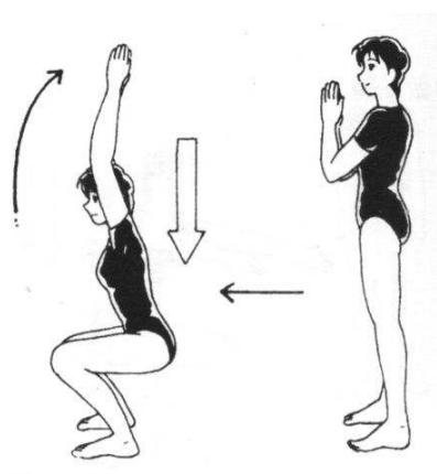
那为什么DNA会开始转录?答案是,有什么东西刺激了它。比如训练,一种施加在肌纤维上的机械外力,也叫机械张力。张,顾名思义,把肌纤维往两边张开、拉开、扯开。
例如在哑铃弯举中,重力作用于哑铃,哑铃把肌纤维往下“扯”,我们自己的骨骼支撑,把肌纤维往上拉,则构成了一个往两边张开的力。

图19:机械张力
机械张力为什么能刺激DNA表达[87][88][89](转录)呢?因为我们有大量的生物感受器,能把外力信号,转变为细胞内的生物信号,这些信号一直传递到DNA上,刺激了DNA的转录,于是我们得到了肌细胞内的蛋白质。
图20:机械刺激与细胞信号
肌肉上能感知机械张力的生物传感器有哪些呢?
例如肋节[90][91][92],它将肌细胞膜与肌原纤维、细胞外基质连接起来,加强肌细胞膜的稳定性和强度,还能感受、侦测到施加于及细胞的外力(例如我们所说的机械张力),将其传导到肌细胞内部,转化为生物信号[16,17];
71整合素[93][94]也是一种横跨细胞膜的受体,它一方面提供连接作用[95],一方面将机械信号从细胞外传递到细胞内。还有磷脂酸(PA)、FAK—粘着斑激酶等也参与机械张力转化为细胞信号的传导传导,就不多说了。

图21:生物传感器
五、训练是如何刺激DNA表达的?答案是细胞信号
生物感受器将细胞信号传递到细胞内,引发一系列细胞信号事件。其中最著名、最核心的细胞信号事件,也被称为PI3K/Akt/mTOR路径[96][97][98][99][100]。还有一些别的相对次要的路径(如ERK),碍于篇幅,我们就不在这里说了。
图22:增肌的核心—mTOR路径
mTOR是我们细胞内一种由2549个氨基酸组成的大型信号蛋白[101]——它既是蛋白质,也发挥信号作用。
mTOR全称“哺乳动物雷帕霉素靶蛋白”,它是哺乳动物调节细胞生长、代谢、蛋白质合成等关键生理过程中的重要蛋白[102]。mTOR实际上以mTORC1(复合物1)和mTORC2(复合物2)的形式在人体内存在[103]。

图23:mTORC1
MTORC1主要通过S6K1和eIF4E的磷酸化,来引发DNA表达,促进蛋白质合成[104]。此外,S6k1也提高mRNA的翻译效率[105]。
对于健身者来说,最典型的激活mTORC1的因素,当然是训练。
训练刺激(机械张力)可激活mTORC1,使其磷酸化[106][107][108][109][110][111];在mTOR的下游,S6激酶[112][113][114]和eIF4E(真核生物起始因子4E)[115][116]随之也被磷酸化(红色方框)。
图24:机械张力传递到DNA,引发DNA表达
磷酸化是自然界一种非常普遍的、对蛋白质进行化学修饰的过程[117]。
蛋白质磷酸化有效地增加了其复杂性,远远超过了基因组所赋予的多样性[118]。
在磷酸激酶的作用下,生物将磷酸基团加在蛋白质或蛋白类中间产物上,从而将蛋白质磷酸化(或者去磷酸化)。经化学修饰后的蛋白质,功能/生物活性会显著不同。
目前已知的磷酸激酶多大500多种,可针对超过20000种蛋白质上的25000个点位进行磷酸化[119][120][121][122];磷酸化决定了在正常/病理状态下生物体对刺激的反应[123]。
S6k1是DNA转录因子[124],名至实归。S6k1被激活后,接下来核糖体蛋白6被磷酸化,从而增加了核糖体蛋白与5'端寡核苷酸(5’-top)mRNA的亲和力,引起了DNA转录[125][126][127],增加蛋白质合成[128][129][130][131]。

图25:S6k1
反过来,如果蛋白质摄入不足,氨基酸/原料不够,则可以导致核糖体蛋白6的“去磷酸化”[132]。已知的诱导蛋白质去磷酸化的酶超过150种[119]。
研究发现,S6k它的磷酸化水平与增肌之间,存在极强的正相关性,r=0.998。下图纵轴是肌肉增加的幅度,横轴是s6k磷酸化水平,用俗话来说,它们几乎成正比。

图26:S6k磷酸化与增肌
六、能激活mTOR路径的,不止有训练
市面上有一种典型的错误观点,认为训练才能增肌,比如下面图上的这种:

图27:错误观点
注意,图中有2个错误:
错误1:认为增肌的原理是损伤修复。这个错误我们在前面已经解析过了,增肌的主要原理是DNA表达而不是损伤。民间认为损伤增肌,主要是因为损伤是来自于训练,训练能激发DNA表达。
错误2:认为训练是增肌的前提。这也是错的,除了训练,营养[133][134][135][136][137][138][139][140][141]和激素[142][143][144][145][146]也都能激发DNA表达,因为他们的分子路径是高度类似的,原理也相同:都是是通过刺激DNA的表达,来得到更多的肌蛋白。
当然,它们的效果程度不同,这或许是因为三者导致的mTOR磷酸化程度不一样。
图28
我们肌肉中的蛋白质就是这么来的:从激活细胞膜上的受体开始,一个个蛋白/酶依次被激活,最后激活DNA转录,然后mRNA翻译为蛋白质。
许多人很难接受“只打药不练就可以长肌肉”,如果他们知道“只吃蛋白粉不训练也能长肌肉”,估计就更无法接受了。但这是客观事实,不以他们的主观意志为转移。
Liu等人在《临床内分泌与代谢杂志》上发表的了一项以10名健康年轻人为对象的研究,对他们注射氨基酸,观察他们肌细胞内的变化(图30),灰柱“AA”是注射氨基酸后,白色是注射前。纵轴是磷酸化水平。

图30
氨基酸注射,导致p70S6K和eIF4E磷酸化,进而增加了核糖体蛋白S6的磷酸化[147]。S6被磷酸化后,促进了一些在蛋白质合成中起重要作用的核糖体蛋白的合成[148][149]。
这些,也就证明了氨基酸注射或摄入能磷酸化4E-BP1、eIF4E、S6,进而引发蛋白合成:换句话说,单纯的吃蛋白质不运动,也会多多少少长一些肌肉。
除了饮食/营养/训练三者,我们还列举了第四种刺激影响转录因子磷酸化的要素:光。
证据表明,光通过刺激SCN(视交叉上核),来影响ERK、或mTOR路径的下游因子S6核糖激酶的磷酸化[152]。具体的,在我们之前的文章中有过叙述。
https://www.zhihu.com/question/44055485/answer/1992536661
参考
1. ^Frauenfelder H; Sligar SG; Wolynes PG The energy landscapes and motions of proteins. Science 1991, 254, 1598–1603.
2. ^Farhath S, He Z, Nakhla T, et al. Pepsin, a marker of gastric contents, is increased in tracheal aspirates from preterm infants who develop bronchopulmonary dysplasia. Pediatrics 2008;121:253–259.
3. ^Stovold R, Forrest IA, Corris PA, et al. Pepsin, a biomarker of gastric aspiration in lung allografts: a putative association with rejection. Am J Respir Crit Care Med 2007;175:1298–1303.
4. ^Crapko M, Kerschner JE, Syring M, Johnston N. Role of extra‐esophageal reflux in chronic otitis media with effusion. Laryngoscope 2007;117:1419–1423.
5. ^Knight J, Lively MO, Johnston N, Dettmar PW, Koufman JA. Sensitive pepsin immunoassay for detection of laryngopharyngeal reflux. Laryngoscope 2005;115:1473–1478.
6. ^Burkhart J.M., Schumbrutzki C., Wortelkamp S., Sickman A., Zahedi R.P. Systematic and quantitative comparison of digest efficiency and specificity reveals the impact of trypsin quality on MS-based proteomics. J. Proteom. 2012;75:1454–1462.
7. ^Olsen J.V., Ong S.E., Mann M. Trypsin cleaves exclusively C-terminal to arginine and lysine residues. Mol. Cell. Proteom. 2004;3:608–614.
8. ^Keil B. Trypsin. Enzymes. 1971;3:249–275.
9. ^Rodriguez J., Gupta N., Smith R.D., Pevzner P.A. Does trypsin cut before proline? J. Proteom. Res. 2008;7:300–305.
10. ^Matthews DM. Protein absorption. J Clin Pathol Suppl (R Coll Pathol) 1971;5:29–40.
11. ^Matthews DM. Intestinal absorption of peptides. Physiol Rev. 1975 Oct;55(4):537–608.
12. ^Mathews DM, Adibi SA. Peptide absorption. Gastroenterology. 1976 Jul;71(1):151–161.
13. ^Adibi SA. Intestinal transport of dipeptides in man: relative importance of hydrolysis and intact absorption. J Clin Invest. 1971 Nov;50(11):2266–2275.
14. ^Adibi SA. The influence of molecular structure of neutral amino acids on their absorption kinetics in the jejunum and ileum of human intestine in vivo. Gastroenterology. 1969 May;56(5):903–913.
15. ^Adibi SA, Gray SJ. Intestinal absorption of essential amino acids in man. Gastroenterology. 1967 May;52(5):837–845.
16. ^Silk DB. Progress report. Peptide absorption in man. Gut. 1974 Jun;15(6):494–501.
17. ^Adibi SA, Morse EL. The number of glycine residues which limits intact absorption of glycine oligopeptides in human jejunum. J Clin Invest. 1977 Nov;60(5):1008–1016.
18. ^Kania RK, Santiago NA, Gray GM. Intestinal surface amino-oligopeptidases. II. Substrate kinetics and topography of the active site. J Biol Chem. 1977 Jul 25;252(14):4929–4934.
19. ^Rayment I, Holden HM, Whittaker M, Yohn CB, Lorenz M, Holmes KC, Milligan RA (1993) Structure of the actin-myosin complex and its implications for muscle contraction. Science 261:58–65.
20. ^Rayment I, Rypniewski WR, Schmidt-B?se K, Smith R, Tomchick DR, Benning MM, Winkelmann DA, Wesenberg G, Holden HM (1993) Three-dimensional structure of myosin subfragment-1: a molecular motor. Science 261:50–58.
21. ^R Dabrowska, W Drabikowski.Molecular basis of muscular contraction.Postepy Biochem. 1973;19(3):343-59.
22. ^Postepy Biochem.The cross-bridge theory. Journal: Physiological 1973;19(3):343-59.
23. ^Thorson J, White DC. Distributed representations for actin-myosin interaction in the oscillatory contraction of muscle. Biophys J. 1969 Mar;9(3):360–390.
24. ^Wakabayashi K, Sugimoto Y, Tanaka H, Ueno Y, Takezawa Y, Amemiya Y. X-ray diffraction evidence for the extensibility of actin and myosin filaments during muscle contraction. Biophys J. 1994 Dec;67(6):2422–2435.
25. ^R D Bremel, A Weber.Cooperation within actin filament in vertebrate skeletal muscle.Nat New Biol. 1972 Jul 26;238(82):97-101.
26. ^Ricarda Haeger , Felipe de Souza Leite , Dilson E Rassier.Sarcomere length non-uniformities dictate force production along the descending limb of the force-length relation.Proc Biol Sci. 2020 Oct 28;287(1937):20202133.
27. ^Dilson E Rassier.Sarcomere mechanics in striated muscles: from molecules to sarcomeres to J Physiol Cell Physiol. 2017 Aug 1;313(2):C134-C145.Epub 2017 May 24.
28. ^Kiisa Nishikawa 1 , Samrat Dutta 2 , Michael DuVall 2 3 , Brent Nelson 4 , Matthew J Gage 5 , Jenna A Monroy 6.Calcium-dependent titin-thin filament interactions in muscle: observations and theory.J Muscle Res Cell Motil. 2020 Mar;41(1):125-139.Epub 2019 Jul 9.
29. ^ B Brenner, E Eisenberg.The mechanism of muscle contraction. Biochemical, mechanical, and structural approaches to elucidate cross-bridge action in muscle.Basic Res Cardiol. 1987;82 Suppl 2:3-16.
30. ^ Maruyama K, Ebashi S. α-Actinin, a new structural protein from striated muscle. II. Action on actin. J Biochem. 1965;58:13–19.
31. ^Honda K, Yamada T, Endo R, Ino Y, Gotoh M, Tsuda H, Yamada Y, Chiba H, Hirohashi S. Actinin-4, a novel actin-bundling protein associated with cell motility and cancer invasion. J Cell Biol. 1998;140:1383–1393.
32. ^Almenar-Queralt A, Lee A, Conley CA, Ribas de Pouplana L, Fowler VM. Identification of a novel tropomodulin isoform, skeletal tropomodulin, that caps actin filament pointed ends in fast skeletal muscle. J Biol Chem 274: 28466–28475, 1999
33. ^Broschat KO, Weber A, Burgess DR. Tropomyosin stabilizes the pointed end of actin filaments by slowing depolymerization. Biochemistry 28: 8501–8506, 1989
34. ^Fowler VM, Sussmann MA, Miller PG, Flucher BE, Daniels MP. Tropomodulin is associated with the free (pointed) ends of the thin filaments in rat skeletal muscle. J Cell Biol 120: 411–420, 1993
35. ^Gokhin DS, Lewis RA, McKeown CR, Nowak RB, Kim NE, Littlefield RS, Lieber RL, Fowler VM. Tropomodulin isoforms regulate thin filament pointed-end capping and skeletal muscle physiology. J Cell Biol 189: 95–109, 2010
36. ^Lau KF, Dill KA. Theory for protein mutability and biogenesis. Proc Natl Acad Sci U S A. 1990 Jan;87(2):638–642
37. ^Tsai CJ; Ma B; Sham YY; Kumar S; Nussinov R Structured disorder and conformational selection. Proteins 2001, 44, 418–427.
38. ^Tzeng SR; Kalodimos CG Protein dynamics and allostery: an NMR view. Curr. Opin. Struct. Biol 2011, 21, 62–67.
39. ^Zhuravlev PI; Papoian GA Protein functional landscapes, dynamics, allostery: a tortuous path towards a universal theoretical framework. Q. Rev. Biophys 2010, 43, 295–332.
40. ^Chan HS, Dill KA. Origins of structure in globular proteins. Proc Natl Acad Sci U S A. 1990 Aug;87(16):6388–6392.
41. ^Woodward CK, Rosenberg A. Studies of hydrogen exchange in proteins. VI. Urea effects on ribonuclease exchange kinetics leading to a general model for hydrogen exchange from folded proteins. J Biol Chem. 1971 Jul 10;246(13):4114–4121.
42. ^Shortle D, Chan HS, Dill KA. Modeling the effects of mutations on the denatured states of proteins. Protein Sci. 1992 Feb;1(2):201–215
43. ^Venyaminov SY; Rajnavolgyi E; Medgyesi GA; Gergely J; Zavodszky P The role of interchain disulphide bridges in the conformational stability of human immunoglobulin G1 subclass. Hydrogen-deuterium exchange studies. Eur. J. Biochem 1976, 67, 81–86.
44. ^Qin H; Lim L; Song J Protein dynamics at Eph receptor-ligand interfaces as revealed by crystallography, NMR and MD simulations. BMC Biophys. 2012,5,2.
45. ^Karplus M; Weaver DL Protein-folding dynamics. Nature 1976, 260, 404–406.
46. ^McCammon JA; Gelin BR; Karplus M Dynamics of folded proteins. Nature 1977, 267, 585–590.
47. ^Karplus M The Levinthal paradox: yesterday and today. Fold. Des 1997, 2, S69–75.
48. ^Dill KA. Dominant forces in protein folding. Biochemistry. 1990 Aug 7;29(31):7133–7155.
49. ^Dill KA, Bromberg S, Yue K, Fiebig KM, Yee DP, Thomas PD, Chan HS. Principles of protein folding--a perspective from simple exact models. Protein Sci. 1995 Apr;4(4):561–602.
50. ^Ackers GK, Doyle ML, Myers D, Daugherty MA. Molecular code for cooperativity in hemoglobin. Science. 1992 Jan 3;255(5040):54–63.
51. ^Alonso DO, Dill KA. Solvent denaturation and stabilization of globular proteins. Biochemistry. 1991 Jun 18;30(24):5974–5985.
52. ^Shi L, Palleros DR, Fink AL. Protein conformational changes induced by 1,1'-bis(4-anilino-5-naphthalenesulfonic acid): preferential binding to the molten globule of DnaK. Biochemistry. 1994 Jun 21;33(24):7536–7546.
53. ^Almenar-Queralt A, Lee A, Conley CA, Ribas de Pouplana L, Fowler VM. Identification of a novel tropomodulin isoform, skeletal tropo-modulin, that caps actin filament pointed ends in fast skeletal muscle. J Biol Chem. 1999;274:28466–28475.
54. ^Cox PR, Zoghbi HY. Sequencing, expression analysis, and mapping of three unique human tropomodulin genes and their mouse orthologs. Genomics. 2000;63:97–107.
55. ^Fowler VM. Identification and purification of a novel Mr 43,000 tropomyosin-binding protein from human erythrocyte membranes. J Biol Chem. 1987;262:12792–12800.
56. ^ Weber A, Pennise CR, Babcock GG, Fowler VM. Tropomodulin caps the pointed ends of actin filaments. J Cell Biol. 1994;127:1627–1635.
57. ^Anfinsen C.B., Haber E., Sela M., White F.H. The kinetics of formation of native ribonuclease during oxidation of the reduced polypeptide chain. Proc. Natl. Acad. Sci. USA. 1961;47:1309–1314.
58. ^Anfinsen C.B. Principles that govern the folding of protein chains. Science. 1973;181:223–230.
59. ^Svetlov M.S., Kommer A., Kolb V.A., Spirin A.S. Effective cotranslational folding of firefly luciferase without chaperones of the Hsp70 family. Protein Sci. 2006;15:242–247.
60. ^Neurath, H. 1989. Proteolytic processing and physiological regulation. Trends Biochem. Sci. 14:268‐271.
61. ^Hirschberg, C.B. and Snider, M.D. 1987. Topography of glycosylation in the rough endoplasmic reticulum and Golgi apparatus. Annu. Rev. Biochem. 56:63‐89.
62. ^ Suttie, J.W. 1985. Vitamin K–dependent carboxylase. Annu. Rev. Biochem. 54:459‐477.
63. ^Kaufman, R.J. , Murtha, P. , Ingolia, D.E. , Yeung, C.Y. , and Kellems, E.R. 1986. b. Selection and amplification of heterologous genes encoding adenosine deaminase in mammalian cells. Proc. Natl.
64. ^Rothman, M.E. 1994. Mechanisms of intracellular protein transport. Nature 372:55‐63.
65. ^Rothman, J.E. and Orci, L. 1992. Molecular dissection of the secretory pathway. Nature 335:409‐415.
66. ^ Mathews M, Korner A. Mammalian cell-free protein synthesis directed by viral ribonucleic acid. Eur J Biochem. 1970 Dec;17(2):328–338.
67. ^Smith AE, Marcker KA, Mathews MB. Translation of RNA from encephalomyocarditis virus in a mammalian cell-free system. Nature. 1970 Jan 10;225(5228):184–187.
68. ^Leader DP, Klein-Bremhaar H, Wool IG. Distribution of initiation factors in cell fractions from mammalian tissues. Biochem Biophys Res Commun. 1972 Jan 14;46(1):215–224.
69. ^Aviv H, Boime I, Leder P. Protein synthesis directed by encephalomyocarditis virus RNA: properties of a transfer RNA-dependent system. Proc Natl Acad Sci U S A. 1971 Sep;68(9):2303–2307.
70. ^GIACOMONI D, SPIEGELMAN S. Origin and biologic individuality of the genetic dictionary. Science. 1962 Dec 21;138(3547):1328–1331.
71. ^BELOZERSKY AN, SPIRIN AS. A correlation between the compositions of deoxyribonucleic and ribonucleic acids. Nature. 1958 Jul 12;182(4628):111–112.
72. ^Marmur J, Lane D. STRAND SEPARATION AND SPECIFIC RECOMBINATION IN DEOXYRIBONUCLEIC ACIDS: BIOLOGICAL STUDIES. Proc Natl Acad Sci U S A. 1960 Apr;46(4):453–461.
73. ^Ycas M, Vincent WS. A RIBONUCLEIC ACID FRACTION FROM YEAST RELATED IN COMPOSITION TO DESOXYRIBONUCLEIC ACID. Proc Natl Acad Sci U S A. 1960 Jun;46(6):804–811.
74. ^Aviv H, Boime I, Leder P. Protein synthesis directed by encephalomyocarditis virus RNA: properties of a transfer RNA-dependent system. Proc Natl Acad Sci U S A. 1971 Sep;68(9):2303–2307.
75. ^Boime I, Aviv H, Leder P. Protein synthesis directed by encephalomyocarditis virus RNA. II. The in vitro synthesis of high molecular weight proteins and elements of the viral capsid. Biochem Biophys Res Commun. 1971 Nov 5;45(3):788–795.
76. ^Heywood SM, Thompson WC. Studies on the formation of the initiation complex in eukaryotes. Biochem Biophys Res Commun. 1971 May 7;43(3):470–475.
77. ^Lucas-Lenard J. Protein biosynthesis. Annu Rev Biochem. 1971;40:409–448.
78. ^Nathans D. Cell-free protein synthesis directed by coliphage MS2 RNA: synthesis of intact viral coat protein and other products. J Mol Biol. 1965 Sep;13(2):521–531.
79. ^McDowell MJ, Joklik WK. An in vitro protein synthesizing system from mouse L fibroblasts infected with reovirus. Virology. 1971 Sep;45(3):724–733.
80. ^H A ITANO.Human hemoglobin.Science.1953 Jan 30;117(3031):89-94.
81. ^Benz EJ Jr, Forget BG. Defect in messenger RNA for human hemoglobin synthesis in beta thalassemia.The Journal of Clinical Investigation, 01 Dec 1971, 50(12):2755-2760
82. ^Stephen Welle 1 , Kirti Bhatt, Carl A Pinkert.Myofibrillar protein synthesis in myostatin-deficient J Physiol Endocrinol Metab. 2006 Mar;290(3):E409-15.
83. ^Neil Kubica 1 , Douglas R Bolster, Peter A Farrell, Scot R Kimball, Leonard S Jefferson.Resistance exercise increases muscle protein synthesis and translation of eukaryotic initiation factor 2Bepsilon mRNA in a mammalian target of rapamycin-dependent manner.2005 Mar 4;280(9):7570-80.
84. ^Dreyer HC, Fugita S, Cadenas JG, Chinkes DL, Volpi E, Rasmussen B (2006) Resistance exercise increases AMPK activity and reduces 4E-BP1 phosphorylation and protein synthesis in human skeletal muscle. J Physiol 576:613–624
85. ^Baar K, Esser K (1999) Phosphorylation of p70S6k correlates with increased skeletal muscle mass following resistance exercise. Am J Physiol 276:C120–C127
86. ^Luisa Gorza; Matteo Sorge; Laura Seclì; Mara Brancaccio.Master Regulators of Muscle Atrophy: Role of Costamere Components.Cells 2021, 10(1), 61.
87. ^Ervasti JM, Ohlendieck K, Kahl SD, Gaver MG, Campbell KP. Deficiency of a glycoprotein component of the dystrophin complex in dystrophic muscle. Nature 1990;345:315–9.
88. ^Clark KA, McElhinny AS, Beckerle MC, Gregorio CC. Striated muscle cytoarchitecture: an intricate web of form and function. Annu Rev Cell Dev Biol 2002;18:637–706.
89. ^Di Mauro D, Gaeta R, Arco A, Miliardi D, Lentini S, Runci M, et al. Distribution of costameric proteins in normal human ventricular and atrial cardiac muscle. Folia Histochem Cytobiol 2009;47(4):605–8.
90. ^Vatta M, Sinagra G, Brunelli L, Faulkner G. Remodeling of dystrophin and sarcomeric Z-band occurs in pediatric cardiomyopathies: a unifying mechanism for force transmission defect. J Cardiovasc Med (Hagerstown) 2009;10(2):149–56.
91. ^Berthier C, Blaineau S. Supramolecular organization of the subsarcolemmal cytoskeleton of adult skeletal muscle fibers. A review. Biol Cell 89: 413–434, 1997.
92. ^Dreyer HC, Fugita S, Cadenas JG, Chinkes DL, Volpi E, Rasmussen B (2006) Resistance exercise increases AMPK activity and reduces 4E-BP1 phosphorylation and protein synthesis in human skeletal muscle. J Physiol 576:613–624
93. ^Kubica N, Bolster DR, Farrell PA, Kimball SR, JeVerson LS (2005) Resistance exercise increases muscle protein synthesis and translation of eukaryotic initiation factor 2B mRNA in a mammalian target of rapamycin-dependent manner. J Biol Chem 280:7570– 7580.
94. ^Atherton PJ, Babraj J, Smith K, Singh J, Rennie MJ, Wackerhage H (2005) Selective activation of AMPK-PGC-1alpha or PKBTSC2-mTOR signaling can explain speciWc adaptive responses to endurance or resistance training-like electrical muscle stimulation. FASEB J 19:786–788
95. ^Nader GA (2005) Molecular determinants of skeletal muscle mass: getting the AKT together. Int J Biochem Cell Biol 37:1985–1996
96. ^Tidball JG (2005) Mechanical signal transduction in skeletal muscle growth and adaptation. J Appl Physiol 98:1900–1908
97. ^Sabers CJ, Martin MM, Brunn GJ et al.Isolation of a Protein Target of the FKBP12-Rapamycin Complex in Mammalian Cells.The Journal of biological chemistry. 1995;270(2):815–22.
98. ^Saxton RA, Sabatini DM. mTOR Signaling in Growth, Metabolism, and Disease. Cell. 2017;168:960–976.
99. ^Kim D-H, Sarbassov DD, Ali SM et al.mTOR Interacts with Raptor to Form a Nutrient-Sensitive Complex that Signals to the Cell Growth Machinery.Cell. 2002;110(2):163–75.
100. ^ Holz MK, Ballif BA, Gygi SP, Blenis J. mTOR and S6K1 mediate assembly of the translation preinitiation complex through dynamic protein interchange and ordered phosphorylation events. Cell. 2005;123:569–580.
101. ^Ma XM, Yoon SO, Richardson CJ, Julich K, Blenis J. SKAR links pre-mRNA splicing to mTOR/S6K1-mediated enhanced translation efficiency of spliced mRNAs. Cell. 2008;133:303–313.
102. ^Bodine SC, Stitt TN, Gonzalez M, Kline WO, Stover GL, Bauerlein R, Zlotchenko E, Scrimgeour A, Lawrence JC, Glass DJ, Yancopoulos GD. Akt/mTOR pathway is a crucial regulator of skeletal muscle hypertrophy and can prevent muscle atrophy in vivo. Nat Cell Biol 3: 1014 – 1019, 2001.
103. ^Goodman CA, Miu MH, Frey JW, Mabrey DM, Lincoln HC, Ge Y, Chen J, Hornberger TA. A phosphatidylinositol 3-kinase/protein kinase B-independent activation of mammalian target of rapamycin signaling is sufficient to induce skeletal muscle hypertrophy. Mol Biol Cell 21: 3258 –3268, 2010.
104. ^Hornberger TA, McLoughlin TJ, Leszczynski JK, Armstrong DD, Jameson RR, Bowen PE, Hwang ES, Hou H, Moustafa ME, Carlson BA, Hatfield DL, Diamond AM, Esser KA. Selenoprotein-deficient transgenic mice exhibit enhanced exercise-induced muscle growth. J Nutr 133: 3091–3097, 2003.
105. ^Hornberger TA, Stuppard R, Conley KE, Fedele MJ, Fiorotto ML, Chin ER, Esser KA. Mechanical stimuli regulate rapamycin-sensitive signaling by a phosphoinositide 3-kinase-, protein kinase B- and growth factor-independent mechanism. Biochem J 380: 795–804, 2004.
106. ^Kubica N, Bolster DR, Farrell PA, Kimball SR, Jefferson LS. Resistance exercise increases muscle protein synthesis and translation of eukaryotic initiation factor 2Bepsilon mRNA in a mammalian target of rapamycin-dependent manner. J Biol Chem 280: 7570 –7580, 2005.
107. ^Miyazaki M, McCarthy JJ, Fedele MJ, Esser KA. Early activation of mTORC1 signalling in response to mechanical overload is independent of phosphoinositide 3-kinase/Akt signalling. J Physiol 589.7: 1831–1846, 2011.
108. ^Pallafacchina, G., Calabria, E., Serrano, A. L., Kalhovde, J. M.,& Schiaffino, S. (2002). A protein kinase B-dependent and rapamycin- sensitive pathway controls skeletal muscle growth but not fiber type specification. Proc. Natl. Acad. Sci. U.S.A.,25.
109. ^Inoki, K., Li, Y., Zhu, T., Wu, J., & Guan, K. L. (2002). TSC2 is phosphorylated and inhibited by Akt and suppresses mTOR signalling.Nat. Cell. Biol., 4, 648–657.
110. ^Hannan, K. M., Thomas, G., & Pearson, R. B. (2003). Activation of S6K1 (p70 ribosomal protein S6 kinase 1) requires an initial calcium-dependent priming event involving formation of a highmolecular-mass signalling complex. Biochem. J., 370, 469–477.
111. ^Kimball SR, Jefferson LS, Fadden P, Haystead TAJ, Lawrence JC 1996 Insulin and diabetes cause reciprocal changes in the association of eIF-4E and PHAS-I in rat skeletal muscle. Am J Physiol 270:C705–C709.
112. ^Pause A, Belsham G, Gingras AC, Donze O, Lin TA, Lawrence JC, Sonenberg N 1994 Insulin-dependent stimulation of protein synthesis by phosphorylation of a regulator of 5-cap function. Nature 371:762–767.
113. ^C S Rubin, O M Rosen.Protein phosphorylation.Annu Rev Biochem. 1975;44:831-87.
114. ^ab Alonso A, Sasin J, Bottini N, Friedberg I, Friedberg I, Osterman A, Godzik A, Hunter T, Dixon J, and Mustelin T (2004). Protein tyrosine phosphatases in the human genome. Cell 117, 699–711.
115. ^Hornbeck PV, Kornhauser JM, Tkachev S, Zhang B, Skrzypek E, Murray B, Latham V, and Sullivan M (2012). PhosphoSitePlus: a comprehensive resource for investigating the structure and function of experimentally determined post-translational modifications in man and mouse. Nucleic Acids Res 40, D261–270.
116. ^ Khoury GA, Baliban RC, and Floudas CA (2011). Proteome-wide post-translational modification statistics: frequency analysis and curation of the swiss-prot database. Sci. Rep 1.
117. ^Manning G, Whyte DB, Martinez R, Hunter T, and Sudarsanam S (2002). The protein kinase complement of the human genome. Science 298, 1912–1934.
118. ^Pawson T, and Scott JD (2005). Protein phosphorylation in signaling--50 years and counting. Trends Biochem. Sci 30, 286–290.
119. ^Satoshi Fujita, Hans C Dreyer, Micah J Drummond, Erin L Glynn, Jerson G Cadenas, Fumiaki Yoshizawa, Elena Volpi, Blake B Rasmussen.Nutrient signalling in the regulation of human muscle protein synthesis.J Physiol. 2007 Jul 15;582(Pt 2):813-23.
120. ^Jefferies H B J, Reinhard C, Kozma S C, Thomas G. Rapamycin selectively represses translation of the 'polypyrimidine tract' mRNA family. Proc Natl Acad Sci USA. 1994;91:4441–4445.
121. ^Jefferies H B J, Thomas G, Thomas G. Elongation factor-1α mRNA is selectively translated following mitogenic stimulation. J Biol Chem. 1994;269:4367–4372.
122. ^Thomas G, Thomas G. Translational control of mRNA expression during the early mitogenic response in Swiss mouse 3T3 cells: identification of specific proteins. J Cell Biol. 1986;103:2137–2144.
123. ^Gruner S., Peter D., Weber R., Wohlbold L., Chung M.Y., Weichenrieder O., Valkov E., Igreja C., Izaurralde E. The structures of eIF4E-eIF4G complexes reveal an extended interface to regulate translation initiation. Mol. Cell. 2016;64:467–479.
124. ^Kubica, N., Bolster, D. R., Farrell, P. A., Kimball, S. R., & Jefferson,L. S. (2005). Resistance exercise increases muscle protein synthesis and translation of eukaryotic initiation factor 2B{epsilon} mRNA in a mammalian target of rapamycin-dependent manner.J. Biol. Chem., 280, 7570–7580.
125. ^Proud CG, Denton RM 1997 Molecular mechanisms for the control of translation by insulin. Biochem J 328:329–341
126. ^Kleijn M, Scheper GC, Voorma HO, Thomas AA 1998 Regulation of translation initiation factors by signal transduction. Eur J Biochem 253:531–544.
127. ^Thomas G, Thomas G. Translational control of mRNA expression during the early mitogenic response in Swiss mouse 3T3 cells: identification of specific proteins. J Cell Biol. 1986;103:2137–2144.
128. ^Karlsson HK, Nilsson PA, Nilsson J, Chibalin AV, Zierath JR, Blomstrand E (2004) Branched-chain amino acids increase p70S6k phosphorylation in human skeletal muscle after resistance exercise. Am J Physiol Endocrinol Metab 287:E1–E7
129. ^Douglas Paddon-Jones 1 , Melinda Sheffield-Moore, Xiao-Jun Zhang, Elena Volpi, Steven E Wolf, Asle Aarsland, Arny A Ferrando, Robert R Wolfe.Amino acid ingestion improves muscle protein synthesis in the young and elderly.Am J Physiol Endocrinol Metab. 2004 Mar;286(3):E321-8.
130. ^K Smith, J M Barua, P W Watt, C M Scrimgeour, M J Rennie.Flooding with L-[1-13C]leucine stimulates human muscle protein incorporation of continuously infused L-[1-13C]valine.Am J Physiol. 1992 Mar;262(3 Pt 1):E372-6.
131. ^Volpi E, Kobayashi H, Sheffield-Moore M, Mittendorfer B, Wolfe RR. Essential amino acids are primarily responsible for the amino acid stimulation of muscle protein anabolism in healthy elderly adults. Am J Clin Nutr. 2003;78:250–8.
132. ^P J Garlick, I Grant.Amino acid infusion increases the sensitivity of muscle protein synthesis in vivo to insulin. Effect of branched-chain amino acids.Biochem J. 1988 Sep 1;254(2):579-84.
133. ^Koopman R, Crombach N, Gijsen AP, et al. Ingestion of a protein hydrolysate is accompanied by an accelerated in vivo digestion and absorption rate when compared with its intact protein. Am. J. Clin. Nutr. 2009; 90:106Y15.
134. ^Jeffery Escobar, Jason W Frank, Agus Suryawan, Hanh V Nguyen, Scot R Kimball, Leonard S Jefferson, Teresa A Davis.Regulation of cardiac and skeletal muscle protein synthesis by individual branched-chain amino acids in neonatal pigs.Am J Physiol Endocrinol Metab. 2006 Apr;290(4):E612-21.
135. ^J C Anthony, T G Anthony, S R Kimball, T C Vary, L S Jefferson.Orally administered leucine stimulates protein synthesis in skeletal muscle of postabsorptive rats in association with increased eIF4F formation.J Nutr. 2000 Feb;130(2):139-45.
相关文章


最新文章
-
中国民间疗法:流行在华佗故乡的有效民间单方
13分钟前 -

诊要经终论第十六之二四时剌法依人气所在而有...
24分钟前 -
肌无力早期的4大症状 中医如何治疗肌无力
44分钟前 -
山西中医世家郭氏秘传肝硬化经验方(详分十种证型,精细解析,深...
57分钟前 -
泥丸脑心命门咏(十五)
1小时前 -
细辛治阳痿早泄
1小时前 -
中医传统保健穴位养生——针灸艾灸治疗坐骨神经痛、腰椎间盘突出...
1小时前 -
此方治疗顽固性甲状腺结节、淋巴结节效果竟如此神奇!
2小时前
热门文章
-

干咳、百日咳、顽痰久咳、肠燥便秘,一个奇效方,对证用药效果好
1周前 -
整椎DIY──滚背脊椎诊断要点
3天前 -
胡潇:马克思时间哲学思想发轫
1周前 -
慢性肾炎效验方
1周前 -
口臭归纳为脾胃湿热与脾虚痰湿两个证型,后者用藿香正气胶囊
5天前 -

黄煌:区区五苓散,能医太多病!(上)
1周前 -
记忆力衰退那是你忘了这味安神益智的药膳
5天前 -

选股的各项优化条件
4天前


有话要说...